News & Topics
- July, 2024
Radiomics score derived from T1-w/T2-w ratio image can predict motor symptom progression in Parkinson’s disease.
Eur Radiol (2024). https://doi.org/10.1007/s00330-024-10886-2
- June, 2024
- May, 2024
- April, 2024
A new paper publishrd on Radiol Phys Technol (IF: 1.6) by Prof. Takatsu.
Takatsu, Y., Ueyama, T., Iwasaki, T. et al.
Simulation of time–intensity curve based on k-space filling in breast dynamic contrast-enhanced three-dimensional magnetic resonance imaging.
Radiol Phys Technol (2024). https://doi.org/10.1007/s12194-024-00793-y
A new paper publishrd on Radiol Phys Technol (IF: 4.4) by Associate Prof. Shiiba.
Shiiba, T., Watanabe, M.
Stability of radiomic features from positron emission tomography images: a phantom study comparing advanced reconstruction algorithms and ordered subset expectation maximization.
Phys Eng Sci Med (2024). https://doi.org/10.1007/s13246-024-01416-x
New under graduate students joined our labs.
Takatus Lab's WEB page has launched.
https://sites.google.com/view/takatsulab-fujita/home
- Feburary, 2024
Takatsu Y, Ohnishi H, Tateyama T, Miyati T.
Usefulness of fat-containing agents: an initial study on estimating fat content for magnetic resonance imaging.
Phys Eng Sci Med. Published online February 20, 2024. doi:10.1007/s13246-023-01372-y
- November, 25–26 2023
- November, 3–4 2023
- July, 2023
- June, 2023
- April, 2023
Mr. Shimozono (M2 student) made an oral presentation at the 79th JSRT.
- April, 2023
- March, 2023
Dependence of apparent diffusion coefficient on slice position in magnetic resonance diffusion imaging.
Takatsu Y, Nakamura M, Suzuki Y, Miyati T. Magn Reson Imaging. 2023;99:41-47. doi:10.1016/j.mri.2023.01.009
- 12 March, 2023
- December, 2022
"Can magnetic resonance imaging after cranioplasty using titanium mesh detect brain tumors?."
Takatsu, Y., Yoshida, R., Yamatani, Y. et al. Phys Eng Sci Med (2022). https://doi.org/10.1007/s13246-022-01200-9
- December, 2022
- April 16, 2022
- April 1, 2022
- March 13, 2022
- June 1, 2021
- April 1, 2021
Introduction
lecular imaging is a diagnostic imaging technique that provides a detailed picture of what is going on in the body at the molecular and cellular levels. While other imaging modalities such as X-rays, computed tomography (CT), and ultrasound provide images of body structures, molecular imaging allows us to see how the body functions and measure chemical and biological processes. Molecular imaging includes diagnostic imaging techniques such as MRI and nuclear medicine, which can help catch diseases in their early stages and pinpoint the exact location of tumors. In molecular imaging, our goal is to develop and evaluate these imaging methods, develop new diagnostic indices by image analysis, and clarify their relationship to prognosis.
Members
Faculty members
Radiological technology
-
 Yasuo Takatsu
(Medical Professor) -
 Seiji Shirakawa
(Associate Professor) -
 Takuro Shiiba
(Associate Professor) -
 Kazuki Takano
(Assistant Professor)
Research Theme
1. Researches on MRI
I studied the physiological processes of the body by using magnetic resonance imaging. With imaging techniques and their resultant clinical applications, I intended to provide information from the analysis and evaluation of the resulting images.
・Development of a new imaging method using SSFP in the female pelvis
・Development of a new evaluation method for liver Gd-EOB-DTPA images
・Study of RF shielding effect of titanium mesh after cranioplasty
・Distortion correction of diffusion-weighted images by non-rigid image registration.
・Dependence of k-space trajectory on time-intensity curve of mammary gland MRI
(Yasuo Takatsu)
2. Development of imaging biomarkers for neurodegenerative diseases
2. Dosimetry in nuclear medicine using Monte Carlo simulation
The development of biomarkers for Alzheimer's disease and Parkinson's disease is an essential issue in a society that aims for long and healthy life. We aim to develop new imaging biomarkers from molecular images such as MRI, PET, and SPECT, in addition to conventional quantitative values. We search for effective image biomarkers for early diagnosis, stratification, and prognosis and develop classification and prognosis models using machine learning.
In addition, we are also working on dose assessment using Monte Carlo simulation in nuclear medicine. (Takuro Shiiba)
3. Development of analyzing method for brain function and structure using MRI and image processing techniques to improve the accuracy for assessment
Magnetic resonance imaging (MRI) allows to show the contrast depending on the condition and behavior of water molecule in tissues, and gives very detailed images of soft tissues like the brain. Our research focuses on developing analysis methods to noninvasively assess of brain function and structure in neurological disorders using MRI, and image processing techniques to improve the accuracy to assess.(Kazuki Takano)
Annual Events
Journal club
Faculty members and graduate students introduce papers on the latest findings.
Research meeting
 Research Meeting
Undergraduate and graduate students will present their research progress.
Graduation Research Presentation
Undergraduate students will present their graduation research, which they have been working on for about six months.
Academic conference presentations
 Presentation on an academic conference
Faculty members and graduate students will present at domestic conferences such as the Japanese Society of Magnetic Resonance Medicine (JSMRM) and the Japanese Society of Radiological Technology (JSRT) and overseas conferences such as the Society of Nuclear Medicine and Molecular Imaging (SNMMI). Undergraduate students are also expected to present their research at domestic and regional conferences.
Photo Gallery
-

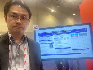
Associate Prof. Shiiba has presented at SNMMI 2024 (Toronto, ON, Canada)
-

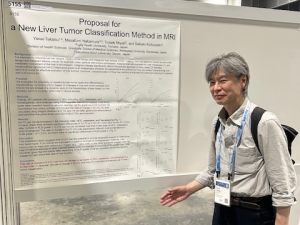
Prof. Takatsu has presented at ISMRM 2024 (Singapore).
-

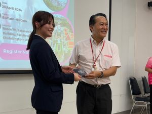
Mrs. Fujita (Shirakawa Lab. M1) has presented at the 1st joint conference of Chulalongkon Univ. and Fujita Health Univ. in Radiological Technology.
-

Mr.Watanabe (Takatsu Lab. B4 student ) presented at CCRT2023 (Fukui, Japan)
-

AAPM2023
-

Welcome party for 2023
-

Graduate ceremony 2022
-

Graduation research presentations
-

Members of 2022 Shiiba-Takano lab
-

Graduate ceremony
-
 Poster presentation on SNMMI2019
-
 SNMMI2019 (Anaheim, USA)
-
 EANM2018 (Düsseldorf, Germany)
Academic Activities
Manuscripts
2024
- Takatsu, Y., Ueyama, T., Iwasaki, T. et al.
Simulation of time–intensity curve based on k-space filling in breast dynamic contrast-enhanced three-dimensional magnetic resonance imaging.
Radiol Phys Technol (2024). https://doi.org/10.1007/s12194-024-00793-y - Shiiba, T., Watanabe, M.Stability of radiomic features from positron emission tomography images: a phantom study comparing advanced reconstruction algorithms and ordered subset expectation maximization. Phys Eng Sci Med (2024). https://doi.org/10.1007/s13246-024-01416-x
- Shimozono, T., Shiiba, T. & Takano, K. Radiomics score derived from T1-w/T2-w ratio image can predict motor symptom progression in Parkinson’s disease. Eur Radiol (2024). https://doi.org/10.1007/s00330-024-10886-2
2023
- Takatsu Y, Nakamura M, Suzuki Y, Miyati T. Dependence of apparent diffusion coefficient on slice position in magnetic resonance diffusion imaging. Magn Reson Imaging. 2023;99:41-47. doi:10.1016/j.mri.2023.01.009
- Naoya Kuga, Takuro Shiiba, Tatsuhiko Sato, Shintaro Hashimoto & Yasuyoshi Kuroiwa. Experimental and computational verifications of the dose calculation accuracy of PHITS for high-energy photon beam therapy, Journal of Nuclear Science and Technology, DOI: 10.1080/00223131.2023.2275737
2022
- Takatsu Y, Nakamura M, Suzuki Y, Miyati T. Dependence of apparent diffusion coefficient on slice position in magnetic resonance diffusion imaging. Magn Reson Imaging. 2023;99:41-47. doi:10.1016/j.mri.2023.01.009
- Takatsu, Y., Yoshida, R., Yamatani, Y. et al. Can magnetic resonance imaging after cranioplasty using titanium mesh detect brain tumors?. Phys Eng Sci Med (2022). https://doi.org/10.1007/s13246-022-01200-9
- Yasuo Takatsu, Masafumi Nakamura, Hajime Sagawa, Yuichi Suzuki, Nobuyuki Mori, Shunichi Motegi, Tosiaki Miyati. Differences in apparent diffusion coefficients between normal brain echo- planar images and turbo spin-echo diffusion-weighted images with distortion correction. 149:110202, 2022.
- Yasuo Takatsu, Kenichirou Yamamura, Yuya Yamatani, Daisuke Takahashi, Rei Yoshida, Masaki Asahara, Michitaka Honda, Tosiaki Miyati. Echo‑planar imaging is superior to fast spin‑echo diffusion‑weighted imaging for cranioplasty using titanium mesh in brain magnetic resonance imaging: a phantom study. Radiological Physics and Technology (in press),
- Shiiba T, Sekikawa Y, Tateoka S, et al. Verification of the effect of acquisition time for SwiftScan on quantitative bone single-photon emission computed tomography using an anthropomorphic phantom. EJNMMI Phys. 2022;9(1):48. Published 2022 Jul 30. doi:10.1186/s40658-022-00477-9
- Shiiba, T., Takano, K., Takaki, A. et al. Dopamine transporter single-photon emission computed tomography-derived radiomics signature for detecting Parkinson’s disease. EJNMMI Res 12, 39 (2022). https://doi.org/10.1186/s13550-022-00910-1
2021
- Yasuo Takatsu, Masafumi Nakamura, Satoshi Kobayashi, Tosiaki Miyati.Prediction of sufficient liver enhancement in the gadoxetate disodium-enhanced hepatobiliary phase by transitional phase images and albumin–bilirubin grade. Magnetic Resonance in Medical Sciences, 20:152-159, 2021,
- Yasuo Takatsu, Tsuyoshi Ueyama, Takahiro Iwasaki, Masaki Asahara, Michitaka Honda, Tosiaki Miyati. Effects of k-space orders on the time-intensity curves in dynamic contrast-enhanced magnetic resonance imaging of the breast based on simulation study. Magnetic Resonance Imaging. 79:85-96, 2021,
- Yasuo Takatsu, Rei Yoshida, Kenichirou Yamamura, Yuya Yamatani, Tsuyoshi Ueyama, Tetsuya Kimura, Yuriko Nohara, Tomohiro Sahara, Kengo Nishiyama, Tosiaki Miyati. Three-dimensional gradient echo sequence is useful for suppressing the radiofrequency shielding effect of a titanium mesh. Magnetic Resonance in Medical Sciences, 20:182-189, 2021,
- Yasuo Takatsu, Masafumi Nakamura, Takanobu Yamashiro, Atsushi Ikemoto, Satoshi Sawa, Masanobu Nakamura, Tosiaki Miyati. Evaluation of contrast and denoising effects related to imaging parameters of compressed sensitivity encoding in contrast‑enhanced magnetic resonance imaging. Radiological Physics and Technology, 14:193-202, 2021,
- Yasuo Takatsu, Masafumi Nakamura, Toshiki Shiozaki, Shoko Narukami, Tosiaki Miyati, Satoshi Kobayashi. Assessment of the cut-off value of quantitative livereportal vein contrast ratio in the hepatobiliary phase of liver MRI. Clinical Radiology, 76:551.e17-551.e24, 2021,
- Eisuke Sato, Kei Fukuzawa, Hiroyuki Takashima, Yuya Yamatani, Yasuo Takatsu, Junichi Hata, Keigo Hikishima, Kenta Miwa. Evaluation of a polyethylene glycol phantom for measuring apparent diffusion coefficients using three 3.0 T MRI systems. Applied Magnetic Resonance, 52:619-631, 2021,
2020
- Shiiba, T, Arimura, Y, Nagano M, Takahashi T, Takaki A. Improvement of classification performance of Parkinson's disease using shape features for machine learning on dopamine transporter single photon emission computed tomography. PLoS ONE, 15(1), e0228289, 2020.
- Takano K, Yamada M. Contrast-enhanced magnetic resonance imaging evidence for the role of astrocytic aquaporin-4 water channels in glymphatic influx and interstitial solute transport. Magnetic Resonance Imaging, 71,11–16, 2020.
- Yasuo Takatsu, Hajime Sagawa, Masafumi Nakamura, Yuichi Suzuki, Tosiaki Miyati. Diffusion-weighted breast magnetic resonance imaging with distortion correction using non-rigid image registration: A clinical study. Radiological Physics and Technology, 13:210–218, 2020,
2019
- Shirakawa, S., K. Tsukamoto, H. Azuma, K. Nakashima, M. Tsujimoto, K. Takano, and M. Yamada. Nonuniform sampling pitch acquisition method in myocardial single-photon emission computed tomography. Nuclear medicine communications, 40(8), 792–801, 2019.
2015
- Yamada M, Takano K, Kawai Y, Kato R. Hemodynamic-based Mapping of Neural Activity in Medetomidine-sedated Rats using a 1.5T Compact Magnetic Resonance Imaging System: A Preliminary Study. Magn Reson Med Sci. 2015;14(3): 243-250.
2010
- Umezawa E, Yoshikawa M, Ohno K, Yoshikawa E, Yamaguchi K. Multi-shelled q-ball imaging: moment-based orientation distribution function. Magnetic Resonance in Medical Sciences 2010;9(3):119-129. DOI: 10.2463/mrms.9.119
2006
- Umezawa E, Yoshikawa M, Yamaguchi K, Ueoku S, Tanaka E. qQ-Space space imaging using small magnetic field gradient. Magnetic Resonance in Medical Sciences 2006;5(4):179-189. DOI: 10.2463/mrms.5.179
Conference
2023
- Yasuo Taktsu. Contrast-Free Tumor Classification in Liver MRI. ICMRI2023, Nov. 3–4, Seoul, Korea.
- Takuya Shimozono and Takuro Shiiba. Magnetic Resonance T1w/T2w Ratio-Based Radiomics Score for Predicting Clinical Motor Outcomes in Parkinson’s Disease. AAPM 65th Annual Meeting, July 23–27, Houston, TX, USA
- Takuro Shiiba. Does radiomics score obtained from dopamine transporter SPECT reflect the severity of Parkinson's disease? SNMMI2023, June 24–27, Chicago IL, USA
2022
-
Yasuo Takatsu, Masafumi Nakamura, Hajime Sagawa, Yuichi Suzuki, Nobuyuki Mori, Shunichi Motegi, Tosiaki Miyati. New reference ADC data for the normal brain with distortion correction in TSE- and EPI-DWI. Joint Annual Meeting ISMRM-ESMEMB ISMRT31st Annual Meetig, 2022.05.07-12 London
-
Yasuo Takatsu, Rei Yoshida, Yuya Yamatani, Mikihisa Kanno, Tosiaki Miyati. Brain Tumor Detectability on the Radio Frequency-Shielding Effect; A Phantom Study. the 10th International Congress on MRI & 27th Annual Scientific Meeting of KSMRM (ICMRI 2022), 2022.11.04-05 Seoul.
2021
- Takuro Shiiba, Yuya Sekikawa, Takuro Shiiba, Shinji Tateoka, Nobutaka Shinohara, Yuuki Inoue, Yasuyoshi Kuroiwa, Yuya Sekikawa, Takashi Tanaka, Yasushi Kihara, Takuroh Imamura. Impact of SwiftScan technique on quantitative bone single photon emission computed tomography. Society of Nuclear Medicine and Molecular Imaging (SNMMI) 2021, June 11-15, 2021, WEB.
- Yuya Sekikawa, Takuro Shiiba, Shinji Tateoka, Nobutaka Shinohara, Yuuki Inoue, Yasuyoshi Kuroiwa, Takashi Tanaka, Yasushi Kihara, Takuroh Imamura. The impact of acquisition time on quantitative bone single photon emission computed tomography using the SwiftScan technique. Society of Nuclear Medicine and Molecular Imaging (SNMMI) 2021, June 11-15, 2021, WEB.
- Yasuo Takatsu, Masafumi Nakamura, Satoshi Kobayashi, Tosiaki MiyatiInvestigation of new assessment method for the liver magnetic resonance imaging. ISMRM & SMRT Virtual Conference & Exhibition. 2021.05.15-20 On line
- Yasuo Takatsu, Kenichiro Yamamura, Yuya Yamatani, Daisuke Takahashi, Rei Yoshida, Masaki Asahara, Michitaka Honda, Tosiaki Miyati. Influence of Radiofrequency-Shielding Effect of a Titanium Mesh in Magnetic Resonance Diffusion-Weighted Imaging. the 9th International Congress on MRI & 26th Annual Scientific Meeting of KSMRM (ICMRI 2021) 2021.11.05-06 Seoul
2020
- Takuro Shiiba, Masahiro Harada, Kaito Ijima. Feasibility of Applying Super-Resolution Techniques Using Deep Learning to SPECT Images. European congress of radiology (ECR) 2021, March 3–7, 2021, WEB.
- Takuro Shiiba, Akihiro Takaki. Feasibility of Improvement of Prediction Performance for Motor Function Prognosis in Parkinson‘s Disease Using Dopamine Transporter SPECT Image Features. 76th Japanese Society of Radiological Technology,May 23-June 17, 2020,WEB.
- Yasuo Takatsu, Hajime Sagawa, Masafumi Nakamura, Yuichi Suzuki, Tosiaki Miyati. Effectiveness of distortion correction for diffusion-weighted imaging using clinical images. ISMRM & SMRT Virtual Conference & Exhibition, 2020.08.08-14 On line
- Yasuo Takatsu, Rei Yoshida, Kinichiro Yamamura, Yuya Yamatani, Tosiaki Miyati. Useful sequence for suppressing the radiofrequency shielding effect of a titanium mesh. ISMRM & SMRT Virtual Conference & Exhibition, 2020.08.08-14 On line
2019
- Takuro Shiiba, Akihiro Takaki. Comparison of diagnostic performance of deep convolutional neural network using fine-tuning and feature extraction on dopamine transporter single photon emission tomography images. Society of Nuclear Medicine and Molecular Imaging (SNMMI) 2019, June 22–25,2019, Anaheim, CA, USA.
- Takuro Shiiba, Yuka Nakamura, Taichi Nakamura, Akihiro Takaki. Development of classification method using automatic shape extraction for dopamine transporter SPECT image. 75th Japanese Society of Radiological Technology,April 11th–14, 2019,Yokohama.
Access
- Access to Fujita Health University ⇒ Fujita Health University Homepage
- Access to Molecular Imaging ⇒ 4th and 5th Floor, University Building #7 and #6

