
Introduction
The "Department of Medical Equipment Engineering", part of the Clinical Collaboration Unit of the Faculty of Medical Sciences, promotes collaboration between the Department of Radiology of the Faculty of Medical Sciences and the Department of Radiology of the University Hospital, with faculty members with scientific and engineering backgrounds and extensive clinical experience working together to promote clinical, research and educational activities. This collaboration covers topics from basic medical equipment to real-time medical care. Our research focuses on diagnostic equipment, from the use of existing equipment to the development of new measurement principles, with the aim of developing radiologists with both clinical and research skills.
Members
Faculty members
Radiological Technology
-
 Yasuki Asada
(Professor)
-
 Hiroshi Kunitomo
(Associate Professor)
-
 Harutoyo Hirano
(Senior Assistant Professor) -
 Masakazu Tsujimoto
(Senior Assistant Professor)
Undergraduate students
ASADA Lab: 5 students; KUNITOMO Lab: 5 students; HIRANO Lab: 6 students
Research Theme
・Research on dosimetry and dose assessment for patients
 Dosimetry using a human equivalent phantom and nanoDot dosimeter
In recent years, it has been reported that medical exposure in Japan is higher than in other countries. The ICRP has recommended the application of diagnostic reference levels (DRLs) as an indicator for optimizing radiation protection. The ICRP recommends the application of the DRLs as an indicator for optimizing radiation protection.
The DRLs were first published in Japan in 2015 and revised in 2020. In preparation for the next revision, we will conduct a nationwide dose survey in general radiography and mammography to clarify the current status of doses received by patients. We are also studying the evaluation and measurement techniques (nanoDot dosimeter) for the entrance surface dose of DRL in general radiography and the glandular dose in mammography. (Yasuki Asada)
・Study on physical image quality assessment method of flat panel detectors (FPDs)
・Studies on optimization of exposure dose based on image quality of digital radiography and angiography
Digital radiography involves radiation exposure, so it is necessary to minimize radiation exposure while still providing useful imaging information for diagnosis and treatment. Our research focuses on elucidating the image characteristics of flat panel detectors (FPDs) used in digital radiography systems (including mammography) and angiography systems to provide better medical images. By elucidating the characteristics of digital imaging systems, we are working to improve examination techniques and optimize image quality and radiation exposure. (Hiroshi Kunitomo)
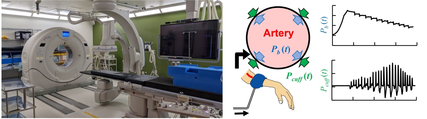
・Study on techniques for measuring human physiological characteristics
・Study on techniques for early diagnosis of atherosclerosis
 Early detection system for atherosclerosis
Humans have highly sophisticated and complex systems for maintaining vital activities such as circulation, respiration, digestion and temperature regulation, which are controlled and processed unconsciously. However, modern state-of-the-art medical technology has not yet been able to reveal the full extent of these complex systems. Unravelling this complex system can lead to an understanding of the mechanisms of disease, quantitative diagnosis of disease and the development of medical devices that can help to establish new treatments for disease. Our laboratory conducts research on the modelling of complex human systems and new engineering technologies useful for medical applications through medical-engineering collaborations, academic-industrial collaborations, and industry-academic collaborations. (Harutoyo Hirano)
・Study on nuclear medicine imaging analysis
Nuclear medicine can image organ and tissue function by administering radioisotopes (RIs). Conventional diagnosis of nuclear medicine images has mainly been based on visual evaluation, however, recent research of nuclear medicine has explored the possibility of predicting treatment efficacy and prognosis by calculating quantitative values and features from the images. Our group is working on various research areas in cooperation with university hospitals in order to conduct technical research for quantitative evaluation and to study the clinical significance by comparing the resultant information obtained from images with histological and clinical diagnoses. (Masakazu Tsujimoto)
Academic Activities
Manuscripts
2024
- Tomonobu Haba, Yusei Nishihara, Yasunori Saito, Takeshi Tomimura, Shuta Ogawa, Kaho Tanabe, Yasuki Asada, Masanao Kobayashi, Shuji Koyama. Estimating organ dose with optimized peak dose index in cone-beam CT scans. Physica Medica. 2024; 118: 103215
- Yuta Matsunaga, Tomonobu Haba, Masanao Kobayashi, Shoichi Suzuki, Yasuki Asada, Koichi Chida. Assessment of fetal radiation exposure in pregnant women undergoing computed tomography and rotational angiography examinations for pelvic trauma. Radiat Prot Dosimetry. 2024; 200(6): 580-587
- Ziqiang Xu, Reiji Anai, Harutoyo Hirano, Zu Soh and Toshio Tsuji. Noninvasive Characterization of Peripheral Sympathetic Activation across Sensory Stimuli Using a Peripheral Arterial Stiffness Index. Frontiers in Physiology, section Computational Physiology and Medicine. 2024; 14: 1294239
- Ziqiang Xu, Zu Soh, Yuta Kurota, Yuya Kimura, Harutoyo Hirano, Takafumi Sasaoka, Atsuo Yoshino and Toshio Tsuji. Neuroimaging-based evidence for sympathetic correlation between brain activity and peripheral vasomotion during pain anticipation. Scientific Reports. 2024; 14, Article number: 3383
2023
- Optimal conversion coefficient from easily measurable dose to effective dose with consideration to radiation quality for posterior-anterior chest radiography Ono, Koji, Asada, Yasuki Biomedical Physics and Engineering Express. 2023; 10(1). doi: 10.1088/2057-1976/ad16c1.
- Evaluation of radiation dose for inferior vena cava filter placement during pregnancy: A comparison of dosimetry and dose calculation software Yuta Matsunaga, Tomonobu Haba, Masanao Kobayashi, Shoichi Suzuki, Yasuki Asada, Koichi Chida J Appl Clin Med Phys. 2023; 24(2): e13884. doi: 10.1002/acm2.13884.
- Genta Tabuchi, Akira Furui, Seiji Hama, Akiko Yanagawa, Koji Shimonaga, Ziqiang Xu, Zu Soh, Harutoyo Hirano & Toshio Tsuji. Motor-cognitive functions required for driving in post-stroke individuals identified via machine-learning analysis. Journal of NeuroEngineering and Rehabilitation. 2023; 20: Article number: 139
- M Tsujimoto, A Fukushima, H Kawai, M Watanabe, S Tanahashi,M Sarai, H Toyama. Volume-based 18F-FDG PET analysis of cardiac sarcoidosis using the descending aorta as a reference tissue. Nuclear Medicine Communications. 2023; 44(5): 390-396
- Taro Okui, Yoshikazu Kobayashi, Madoka Isomura, Masakazu Tsujimoto, Koji Satoh, Hiroshi Toyama. Histopathological findings affect quantitative values of single photon emission computed tomography in patients with antiresorptive agent-related osteonecrosis of the jaws. Fujita medical journal. 2023; 9(3): 186-193
2022
- Yasuki Asada, Honoka Inagaki, Kaito Iwase, Mio Taniguchi, Yuya Nagake, Miuna Hayashi. Measurement of conversion factor into mean glandular dose in mammography using OSL dosimeters. Open Journal of Medical Sciences. 2022; 2(1), 9-16.
- Masanao Kobayashi, Yusei Nishihara, Tomonobu Haba, Yuta Matsunaga, Kazuyuki Minami, Yasuki Asada. SIZE-SPECIFIC DOSE ESTIMATES IN FETAL COMPUTED TOMOGRAPHY. Radiat Prot Dosimetry. 2022; 198(6): 339-348.
- Masanao Kobayashi, Yusei Nishihara, Tomonobu Haba, Yuta Matsunaga, Yasuki Asada, Shigeki Kobayashi. SIZE-specific dose estimate for lower-limb CT. Phys Eng Sci Med. 2022; 45(4): 1183-1191.
- Hideki Shibata, Kosuke Matsubara, Yasuki Asada, Akihiro Takemura, Isao Kozawa. Physical and visual evaluations of CT image quality of large low-contrast objects with visual model-based iterative reconstruction technique: a phantom study. Phys Eng Sci Med. 2023; 46(1): 141-150.
- Yuta Matsunaga 1 2, Tomonobu Haba 3, Masanao Kobayashi 3, Shoichi Suzuki 3, Yasuki Asada 3, Koichi Chida 2 Evaluation of radiation dose for inferior vena cava filter placement during pregnancy: A comparison of dosimetry and dose calculation software. J Appl Clin Med Phys. 2023; 24(2): e13884.
- Kan H, Tsuchiya T, Yamada M, Kunitomo H, Kasai H, Shibamoto Y. Delineation of prostatic calcification using quantitative susceptibility mapping: Spatial accuracy for magnetic resonance-only radiotherapy planning. J Appl Clin Med Phys. 2022 Feb;23(2):e13469. doi: 10.1002/acm2.13469. Epub 2021 Nov 2. PMID: 34726833; PMCID: PMC8833270.
- Kan H, Uchida Y, Ueki Y, Arai N, Tsubokura S, Kunitomo H, Kasai H, Aoyama K, Matsukawa N, Shibamoto Y. R2* relaxometry analysis for mapping of white matter alteration in Parkinson's disease with mild cognitive impairment. Neuroimage Clin. 2022;33:102938. doi: 10.1016/j.nicl.2022.102938. Epub 2022 Jan 4. PMID: 34998126; PMCID: PMC8741619.
- Ryuki Shigemasu, Yuki Teraoka, Satoshi Ota, Harutoyo Hirano, Keita Yasutomi, Shoji Kawahito, Masato Futagawa. Development of a Current Injection—Type Impedance Measurement System for Monitoring Soil Water Content and Ion Concentration. Sensors. 2022; 22(9): 3509.
- Seiya Tanaka, Satoshi Ota, Harutoyo Hirano, Masato Futagawa, Yasushi Takemura. Evaluation of Harmonic Signals Derived From Multiple Spatially Separated Samples for Magnetic Particle Imaging. IEEE Transactions on Magnetics. 2022; 58(8): 1-5, Art no. 6501005.
- Taro Okui, Yoshikazu Kobayashi, Masakazu Tsujimoto, Koji Satoh, Hiroshi Toyama, and Koichiro Matsuo. Correction to: Quantitative evaluation of anti-resorptive agent-related osteonecrosis of the jaw using bone single photon emission computed tomography in clinical settings: relationship between clinical stage and imaging. Ann Nucl Med. 2022; 36(7): 693.
- T Okui, Y Kobayashi, M Isomura, M Tsujimoto, K Satoh, H Toyama. Histopathological findings affect quantitative values of single photon emission computed tomography in patients with antiresorptive agent-related osteonecrosis of the jaws. Fujita Medical Journal, 2023; 9(3), 186-193
2021
- Sakai K.,Yamada Y.,Yoshida K.,Yoshinaga S.,Sato K.,Ogata H.,Iwasaki T.,Kudo S.,Asada Y., Kawaguchi I., Haeno H., Sasaki M.; Conclusions and suggestions on low-dose and low-dose rate radiation risk estimation methodology, J. Radiat. Prot. Res. 2021; 46(1): 14 – 23
- Matsunaga Y., Haba T.,Kobayashi M.,Suzuki S.,Asada Y.,Chida K.; Fetal radiation dose of four tube voltages in abdominal CT examinations during pregnancy: A phantom study, J. Appl .Clin. Med. Phys. 2021; 22(2): 178 – 184
- Nobuhiro Oda, Yoshito Tabata, Masayoshi Mizuta, Yasuki Asada, Tsutomu Nakano, Tatsunori Hara, Yoshiyuki Kurokawa, Takatoshi Aoki, Shuzo Uehara. Optimal Beam Quality in Chest Radiography Using CsI-flat Panel Detector for Detection of Pulmonary Nodules. Jpn J Radiol Tech. 2021; 77(4): 335-343.
- Yuta Matsunaga, Tomonobu Haba, Masanao Kobayashi, Shoichi Suzuki, Yasuki Asada, Koichi Chida. Novel pregnant model phantoms for measurement of foetal radiation dose in x-ray examinations. J Radiol Prot. 2021 Aug 19;41(3): PMID: 34233314.
- Akihiro Arimoto, Yasuki Asada. Investigation of backscatter factor in medical radiography using anthropomorphic phantom by optically stimulated luminescence dosimeter. Biomed Phys Eng Express. 2021; 7(6): PMID: 34438389
- Shinohara N, Akiyama S, Ito T, Okada S, Chiba Y, Negishi T, Hirofuji Y, Kunitomo H. Examination of the Quality Control Items for Digital Breast Tomosynthesis System in Japan. Nihon Hoshasen Gijutsu Gakkai Zasshi. 2021; 77(5): 478-486. Japanese. doi: 10.6009/jjrt.2021_JSRT_77.5.478. PMID: 34011791.
- Kawashima H, Ichikawa K, Kunitomo H. Relationship between Radiation Quality and Image Quality in Digital Chest Radiography: Validation Study Using Human Soft Tissue-equivalent Phantom. Nihon Hoshasen Gijutsu Gakkai Zasshi. 2021; 77(3): 255-262. Japanese. doi: 10.6009/jjrt.2021_JSRT_77.3.255. PMID: 33746173.
- Seiichi Ohkawara, Kentaro Miura, Harutoyo Hirano, Satoshi Ota, Masato Futagawa. Evaluation of a Hydrogen Signal Detection Method Using a Compact NMR Sensor for the Measurement of Ion Concentrations in Culture Medium. IEEJ Transactions on Sensors and Micromachines. 2021; 141(11): 367-372.
- Satoshi Ota, Seiichi Ohkawara, Harutoyo Hirano, Masato Futagawa, Yasushi Takemura. Empirical and simulated evaluations of easy-axis dynamics of magnetic nanoparticles based on their magnetization response in alternating magnetic field. Journal of Magnetism and Magnetic Materials. 2021; 539: Article number 168354.
- Satoshi Kamiya, Ryuji Nakamura, Noboru Saeki, Takashi Kondo, Hirotsugu Miyoshi, Soushi Narasaki, Atsushi Morio, Masashi Kawamoto, Harutoyo Hirano, Toshio Tsuji, Yasuo M. Tsutsumi. Prediction of blood pressure change during surgical incision under opioid analgesia using sympathetic response evoking threshold. Scientific Reports. 2021; 11: Article number 9558. Toshifumi Muneyasu, Harutoyo Hirano, Akira Furui, Zu Soh, Ryuji Nakamura, Noboru Saeki, Yoshiyuki Okada, Masashi Kawamoto, Masao Yoshizumi, Atsuo Yoshino, Takafumi Sasaoka, Shigeto Yamawaki, Toshio Tsuji. Cardiorespiratory synchronisation and systolic blood pressure correlation of peripheral arterial stiffness during endoscopic thoracic sympathectomy. Scientific Reports. 2021; 11: Article number 5966.
- Toshio Tsuji, Fumiya Arikuni, Takafumi Sasaoka, Shin Suyama, Takashi Akiyoshi, Zu Soh, Harutoyo Hirano, Ryuji Nakamura, Noboru Saeki, Masashi Kawamoto, Masao Yoshizumi, Atsuo Yoshino, Shigeto Yamawaki. Peripheral arterial stiffness during electrocutaneous stimulation is positively correlated with pain-related brain activity and subjective pain intensity: an fMRI study. Scientific Reports. 2021; 11: Article number 4425.
- Masakazu Tsujimoto, Atsushi Teramoto, Masakazu Dosho, Shingo Tanahashi, Ayami Fukushima, Seiichiro Ota, Yoshitaka Inui, Ryo Matsukiyo, Yuuki Obama, Hiroshi Toyama. Automated classification of increased uptake regions in bone single-photon emission computed tomography/computed tomography images using three-dimensional deep convolutional neural network. Nucl Med Commun. 2021; 42(8): 877-883.
- Yoshikazu Kobayashi, Taro Okui, Masakazu Tsujimoto, Hirotaka Ikeda, Koji Satoh, Daisuke Kanamori, Naoko Fujii, Hiroshi Toyama, Koichiro Matsuo. Effect of morphological findings in computed tomography on the quantitative values in single-photon emission computed tomography for patients with antiresorptive agent-related osteonecrosis of the jaw: a cross-sectional study. Ann Nucl Med. 2021; 35(7): 853-860.
- Masakazu Tsujimoto, Seiji Shirakawa, Masanori Watanabe, Atsushi Teramoto, Masaki Uno, Seiichiro Ota, Ryo Matsukiyo, Taro Okui, Yoshikazu Kobayashi, Hiroshi Toyama. Two-versus three-dimensional regions of interest for quantifying SPECT-CT images. Phys Eng Sci Med. 2021; 44(2): 365-375.
- Shingo Tanahashi, Masakazu Tsujimoto, Tomoyuki Kurose, Masaki Uno, Masayoshi Sarai, Hideki Kawai, Hiroshi Toyama. Examination of quantitative analysis method for cardiac amyloidosis by 99mTc-PYP scintigraphy. Rinsho Hoshasen 2021; 66(9): 921-928.
- Yuya Onishi, Atsushi Teramoto, Masakazu Tsujimoto, Tetsuya Tsukamoto, Kuniaki Saito, Hiroshi Toyama, Kazuyoshi Imaizumi, Hiroshi Fujita. Investigation of pulmonary nodule classification using multi-scale residual network enhanced with 3DGAN-synthesized volumes. Radiol Phys Technol. 2020; 13(2): 160-169.
Conference
2022
- Hiroshi Kunitomo, “Physical Image Quality Evaluation in Digital Radiography”, The 78th Annual Meeting of the Japanese Society of Radiological Technology, PACIFICO YOKOHAMA and On demand meeting, Apr 2022.
- Harutoyo Hirano, Haruki Goto, Satoshi Ota, Masato Futagawa, Yu Hashimoto, Haruki Hashimoto, Shinji Kishimoto, Nozomu Oda, Masato Kajikawa, Tatsuya Maruhashi, and Yukihito Higashi, “Convolutional Neural Network-Based Assessment Method for Atherosclerosis Using Earlobe Crease Images”, Proceedings of 2022 44th Annual International Conference of the IEEE Engineering in Medicine & Biology Society, p. 2882, Glasgow, UK, July 2022.
- Seiya Tanaka, Harutoyo Hirano, Masato Futagawa, Yasushi Takemura, Satoshi Ota, “Evaluation of harmonic signals derived from separately located multiple samples for magnetic particle imaging”, The 15th Joint MMM-INTERMAG Conference, IPB-17, New Orleans, LA/ondemand, Jan 2022.
- Keita Honda, Kosuke Shimizu, Harutoyo Hirano, Masato Futagawa, Yasushi Takemura, Satoshi Ota, “Investigation of magnetic relaxation of intratumor magnetic nanoparticles for hyperthermia”, The 15th Joint MMM-INTERMAG Conference, IPE-21, New Orleans, LA/ondemand, Jan 2022.
- Kentaro Miura, Seiichi Ohkawara, Harutoyo Hirano, Satoshi Ota, Masato Futagawa, “Examination of a Hydrogen Ion Micro-signal Detection Method Using a Compact Nuclear Magnetic Resonance Sensor for Monitor Supply and Drainage Solutions”, 2022 IEEJ Annual Meeting, 3-161, Online, March 2022.
- Seiya Tanaka, Harutoyo Hirano, Masato Futagawa, Yasushi Takemura, and Satoshi Ota, “Two-dimensional imaging of magnetic nanoparticles under pulsed magnetic field”, Magnetics meeting of IEEJ Fundamentals and Materials (Society A),MAG-22-005, Kanazawa/Online, Jan 2022.
2021
- Takuma Yamada, Harutoyo Hirano, Satoshi Ota, and Masato Futagawa, “Effort to Realize Two-Dimensional Imaging of Soil Moisture Contents Using Machine Learning”, Chemical Sensor Meeting of IEEJ Sensors and Micromachines (Socierty E), CHS-21-032, Saitama/Online, Dec 2021.
- S. Tanaka, H. Hirano, M. Futagawa, Y. Takemura, S. Ota, “Imaging technique of magnetic nanoparticles using pulse magnetic field”, 45th Annual Meeting of MAGNETICS in Japan, 31aA-12, Online, Aug 2021.
- Kentaro Miura, Seiichi Ohkawara , Harutoyo Hirano , Satoshi Ota , Masato Futagawa, “Examination of a Ion Micro-signal Detection Method Using a Compact Nuclear Magnetic Resonance Sensor to Monitor Supply and Drainage Solutions”,Annual Meeting of IEEJ Sensor and Micromachines Society, CHS-21-020, Online July 2021. .
- Haruki Sato, Kisho Sakamoto, Satoshi Ota, Harutoyo Hirano, and Masato Futagawa, “Study of a Soil Water Content Measurement Method by Heat Using a heat Thermoelectric Module”,Annual Meeting of IEEJ Sensor and Micromachines Society, CHS-21-021, Online July 2021.
Access
- Access to Fujita Health University ⇒ Fujita Health University Homepage
- Access to Medical Equipment Engineering
408 and 409, 4th floor, University Building 7